By Bobbie Welty and AI
•
September 26, 2024
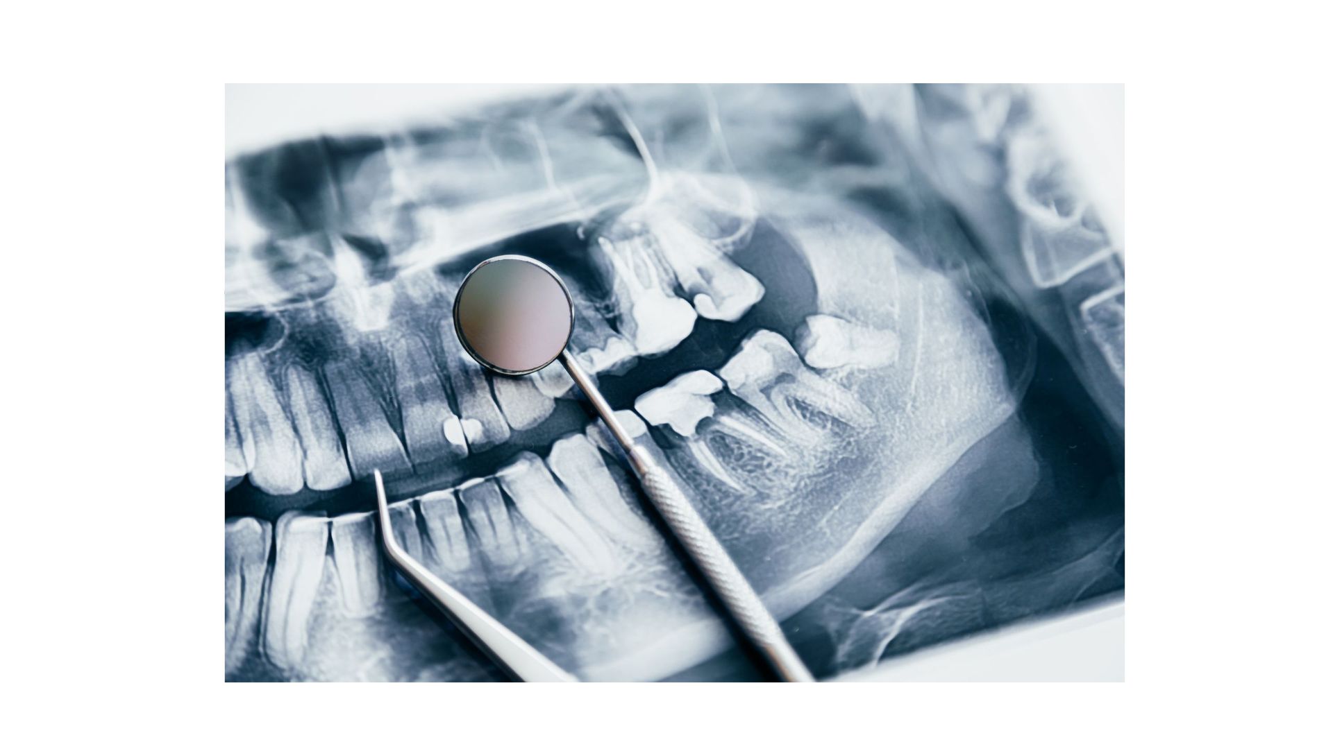
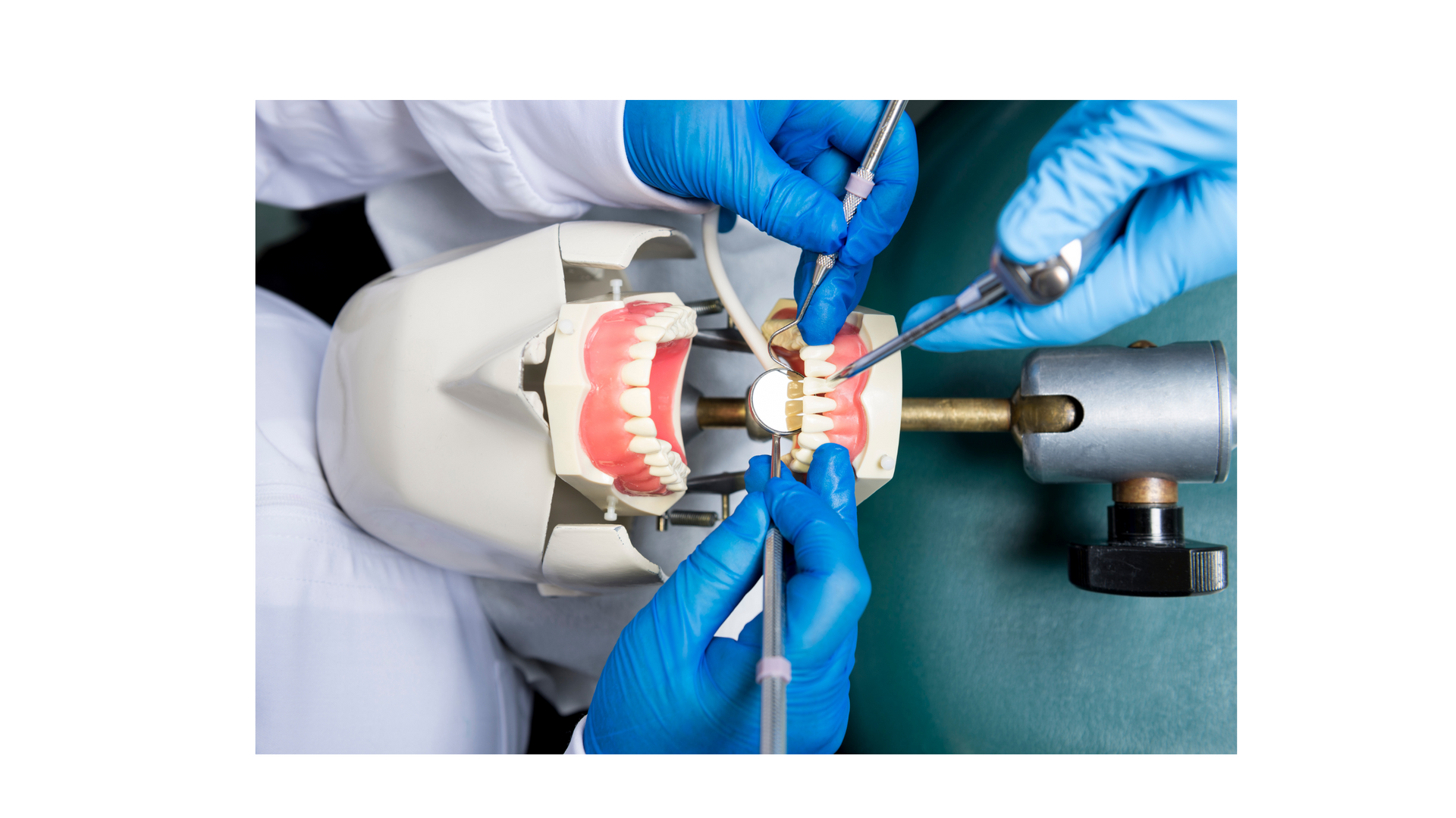
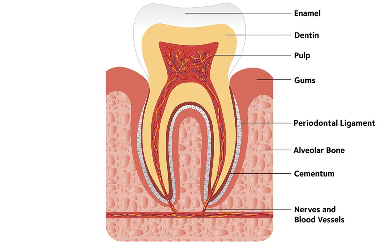
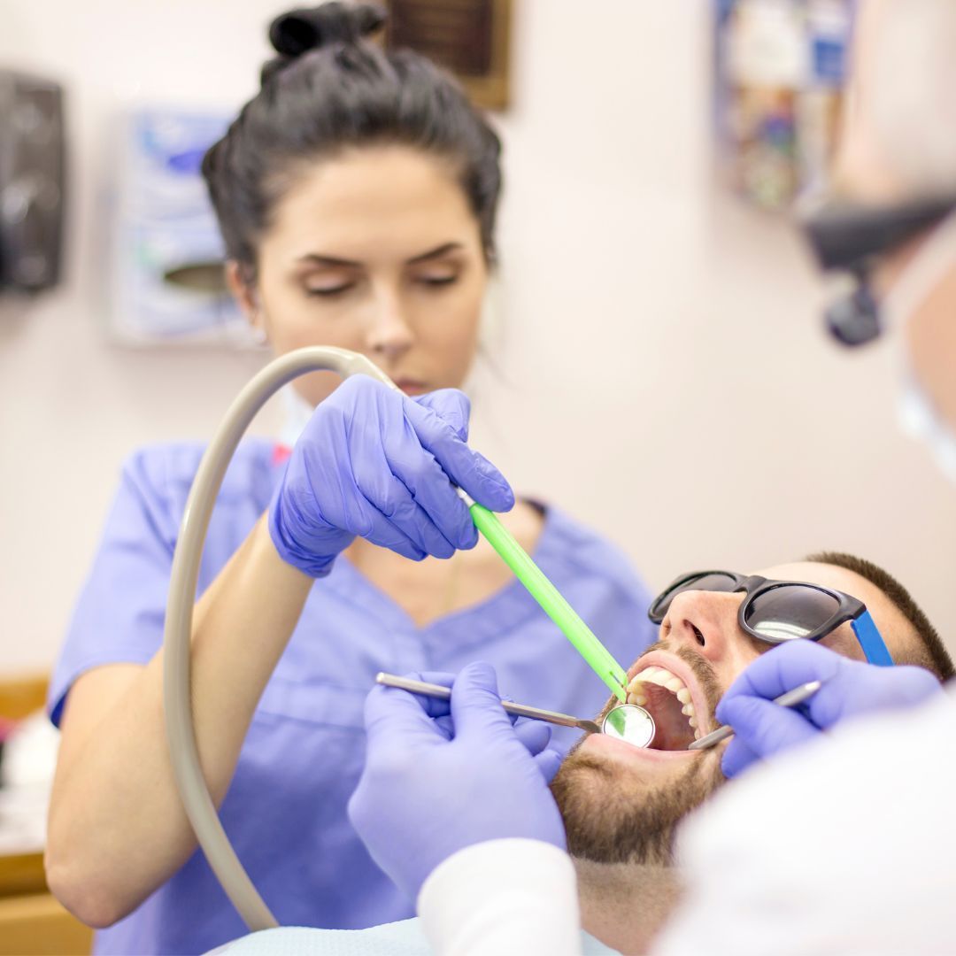
**Understanding the Surfaces and Types of Teeth: A Guide to Your Smile** When we think about teeth, we often focus on how they look or how they function, but did you know that each tooth has a specific role in maintaining oral health? From chewing food to aiding in speech, our teeth are truly fascinating structures. In this post, we’ll dive into the different surfaces of the teeth and explore the various types of teeth in our mouths. **Tooth Surfaces: A Breakdown** Each tooth has multiple surfaces, and understanding them is key to knowing how your dentist evaluates your oral health. 1. **Occlusal Surface** This is the chewing surface of the premolars and molars. These teeth have cusps (pointed parts) that help grind and mash food during chewing. 2. **Buccal Surface** Found on the outside surface of the posterior teeth (molars and premolars), this surface faces the inside of the cheek. The word "buccal" comes from "bucca," which means cheek in Latin. 3. **Lingual Surface** This surface refers to the side of the tooth that faces the tongue. For upper teeth, it's also called the palatal surface because it faces the roof of the mouth, or palate. 4. **Mesial Surface** The mesial surface is the side of the tooth that's closest to the front of the mouth or midline. Think of it as the side of the tooth that touches the adjacent tooth closer to the front. 5. **Distal Surface** The opposite of the mesial surface, the distal surface faces the back of the mouth and is farthest from the midline. It’s the part of the tooth that touches the adjacent tooth closer to the back of the mouth. 6. **Incisal Surface** Found on the anterior teeth (incisors and canines), this surface is the biting edge of the tooth that helps cut food. # **Types of Teeth: What Role Do They Play?** In a healthy adult mouth, there are 32 teeth, and they all play a unique role. Here’s a look at the different types of teeth and their specific functions: 1. **Incisors** The four front teeth on the top and bottom are called incisors. They’re sharp and designed for cutting food. We use these teeth to bite into food like apples and carrots. 2. **Canines** Also known as cuspids, the canines are located next to the incisors. They have a pointed shape designed for tearing food. Canines are often the sharpest teeth in the mouth. 3. **Premolars** Behind the canines are the premolars, also called bicuspids. These teeth have a flat surface with ridges, making them ideal for crushing and tearing food before it reaches the molars. 4. **Molars** Molars are the largest teeth located at the back of the mouth. With their broad, flat surfaces, molars are perfect for grinding food into smaller pieces, making it easier to swallow and digest. 5. **Third Molars (Wisdom Teeth)** The third set of molars, commonly known as wisdom teeth, typically appear in late adolescence or early adulthood. However, not everyone gets their wisdom teeth, and some need to have them removed if they cause overcrowding or other issues. # **Why Is Understanding Tooth Structure Important?** Knowing the different types of teeth and their surfaces can help you understand why dentists focus on particular areas during checkups. For example, occlusal surfaces are prone to decay because they have grooves where food and bacteria can get trapped, while buccal and lingual surfaces can easily accumulate plaque. By taking care of all the surfaces of your teeth through proper brushing and flossing techniques, you can maintain a healthy, radiant smile! I hope this provides a clear understanding of the anatomy of your teeth and the role each part plays. If you have questions or want to learn more about keeping your smile bright, leave a comment below! What do you think? Would you like to tweak or add anything to this draft?Writing a blog on the surfaces and types of teeth can be both informative and engaging for your readers, especially those curious about dental care or aspiring dental professionals. Here's a draft to help get you started: **Understanding the Surfaces and Types of Teeth: A Guide to Your Smile** When we think about teeth, we often focus on how they look or how they function, but did you know that each tooth has a specific role in maintaining oral health? From chewing food to aiding in speech, our teeth are truly fascinating structures. In this post, we’ll dive into the different surfaces of the teeth and explore the various types of teeth in our mouths. # **Tooth Surfaces: A Breakdown** Each tooth has multiple surfaces, and understanding them is key to knowing how your dentist evaluates your oral health. 1. **Occlusal Surface** This is the chewing surface of the premolars and molars. These teeth have cusps (pointed parts) that help grind and mash food during chewing. 2. **Buccal Surface** Found on the outside surface of the posterior teeth (molars and premolars), this surface faces the inside of the cheek. The word "buccal" comes from "bucca," which means cheek in Latin. 3. **Lingual Surface** This surface refers to the side of the tooth that faces the tongue. For upper teeth, it's also called the palatal surface because it faces the roof of the mouth, or palate. 4. **Mesial Surface** The mesial surface is the side of the tooth that's closest to the front of the mouth or midline. Think of it as the side of the tooth that touches the adjacent tooth closer to the front. 5. **Distal Surface** The opposite of the mesial surface, the distal surface faces the back of the mouth and is farthest from the midline. It’s the part of the tooth that touches the adjacent tooth closer to the back of the mouth. 6. **Incisal Surface** Found on the anterior teeth (incisors and canines), this surface is the biting edge of the tooth that helps cut food. **Types of Teeth: What Role Do They Play?** In a healthy adult mouth, there are 32 teeth, and they all play a unique role. Here’s a look at the different types of teeth and their specific functions: 1. **Incisors** The four front teeth on the top and bottom are called incisors. They’re sharp and designed for cutting food. We use these teeth to bite into food like apples and carrots. 2. **Canines** Also known as cuspids, the canines are located next to the incisors. They have a pointed shape designed for tearing food. Canines are often the sharpest teeth in the mouth. 3. **Premolars** Behind the canines are the premolars, also called bicuspids. These teeth have a flat surface with ridges, making them ideal for crushing and tearing food before it reaches the molars. 4. **Molars** Molars are the largest teeth located at the back of the mouth. With their broad, flat surfaces, molars are perfect for grinding food into smaller pieces, making it easier to swallow and digest. 5. **Third Molars (Wisdom Teeth)** The third set of molars, commonly known as wisdom teeth, typically appear in late adolescence or early adulthood. However, not everyone gets their wisdom teeth, and some need to have them removed if they cause overcrowding or other issues. **Why Is Understanding Tooth Structure Important?** Knowing the different types of teeth and their surfaces can help you understand why dentists focus on particular areas during checkups. For example, occlusal surfaces are prone to decay because they have grooves where food and bacteria can get trapped, while buccal and lingual surfaces can easily accumulate plaque. By taking care of all the surfaces of your teeth through proper brushing and flossing techniques, you can maintain a healthy, radiant smile! I hope this provides a clear understanding of the anatomy of your teeth and the role each part plays. If you have questions or want to learn more about keeping your smile bright, leave a comment below!